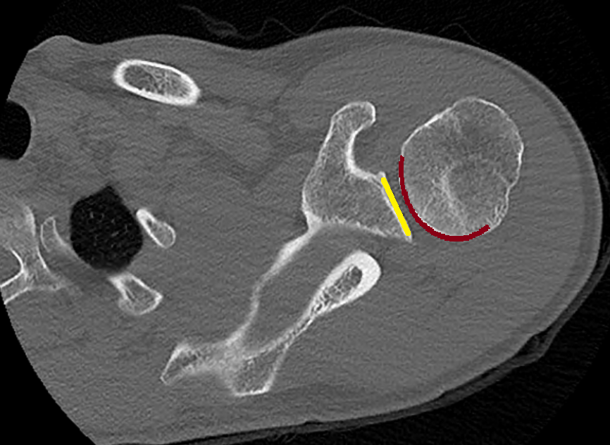
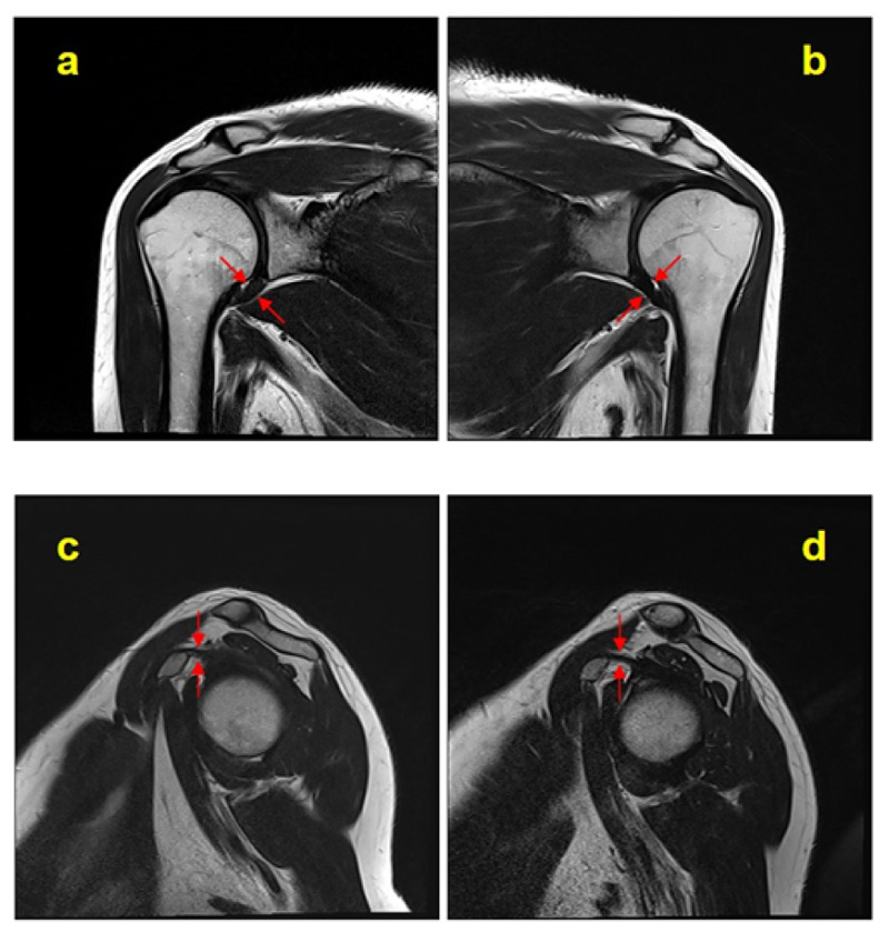
Typical magnetic resonance imaging scan showing the coracohumeral
Por um escritor misterioso
Descrição
Glenohumeral-Instability – OrthoPaedia

BIR Publications
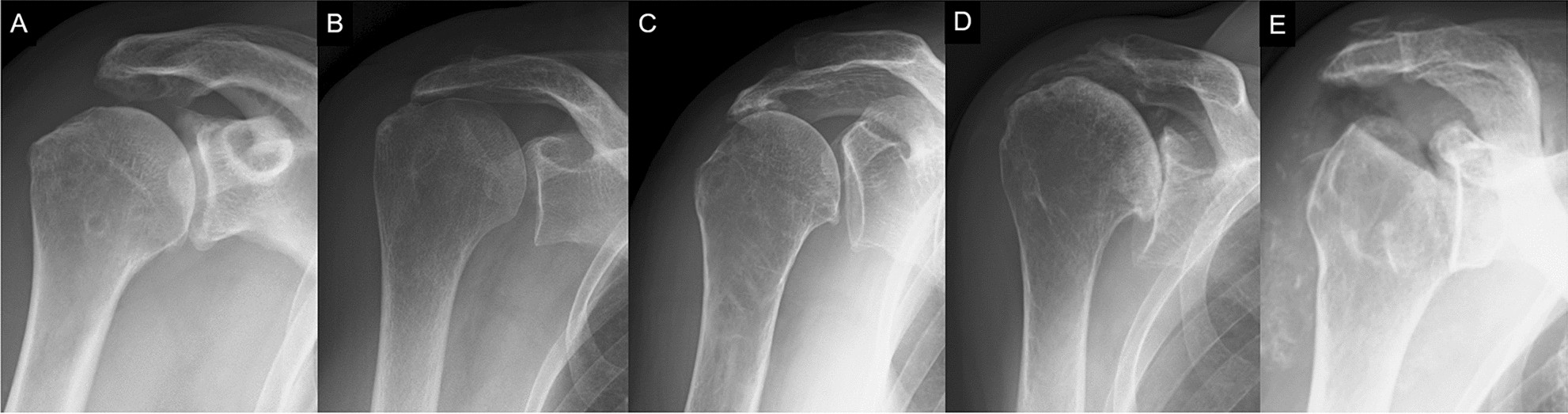
Risk factors of radiographic severity of massive rotator cuff tear
Miscellaneous Conditions of the Shoulder

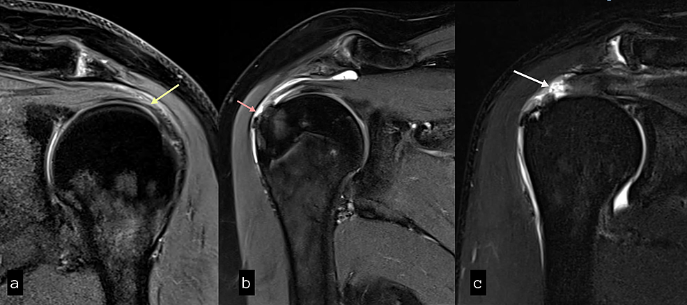
Magnetic resonance imaging of the shoulder
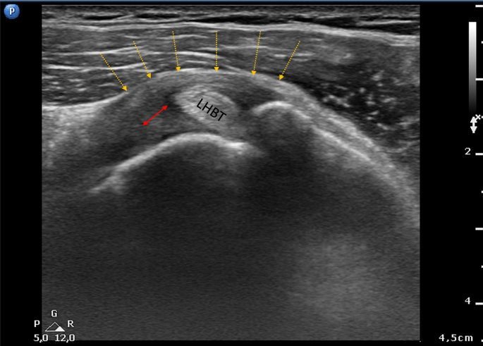
Ultrasound Features of Adhesive Capsulitis
Contrast-enhanced Magnetic Resonance Imaging Revealing the Joint Capsule Pathology of a Refractory Frozen Shoulder

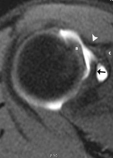
Beyond the Cuff: MR Imaging of Labroligamentous Injuries in the Athletic Shoulder
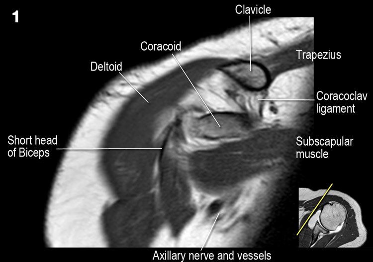
The Radiology Assistant : Shoulder Anatomy and Variants on MRI
Cureus, Role of Magnetic Resonance Imaging in the Evaluation of Rotator Cuff Tears

Pain related to rotator cuff abnormalities: MRI findings without clinical significance - Bencardino - 2010 - Journal of Magnetic Resonance Imaging - Wiley Online Library
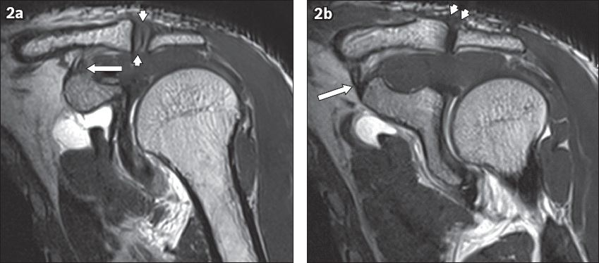
Spectrum of lesions of the acromioclavicular joint: imaging features
de
por adulto (o preço varia de acordo com o tamanho do grupo)