Radiological identification and analysis of soft tissue musculoskeletal calcifications, Insights into Imaging
Por um escritor misterioso
Descrição
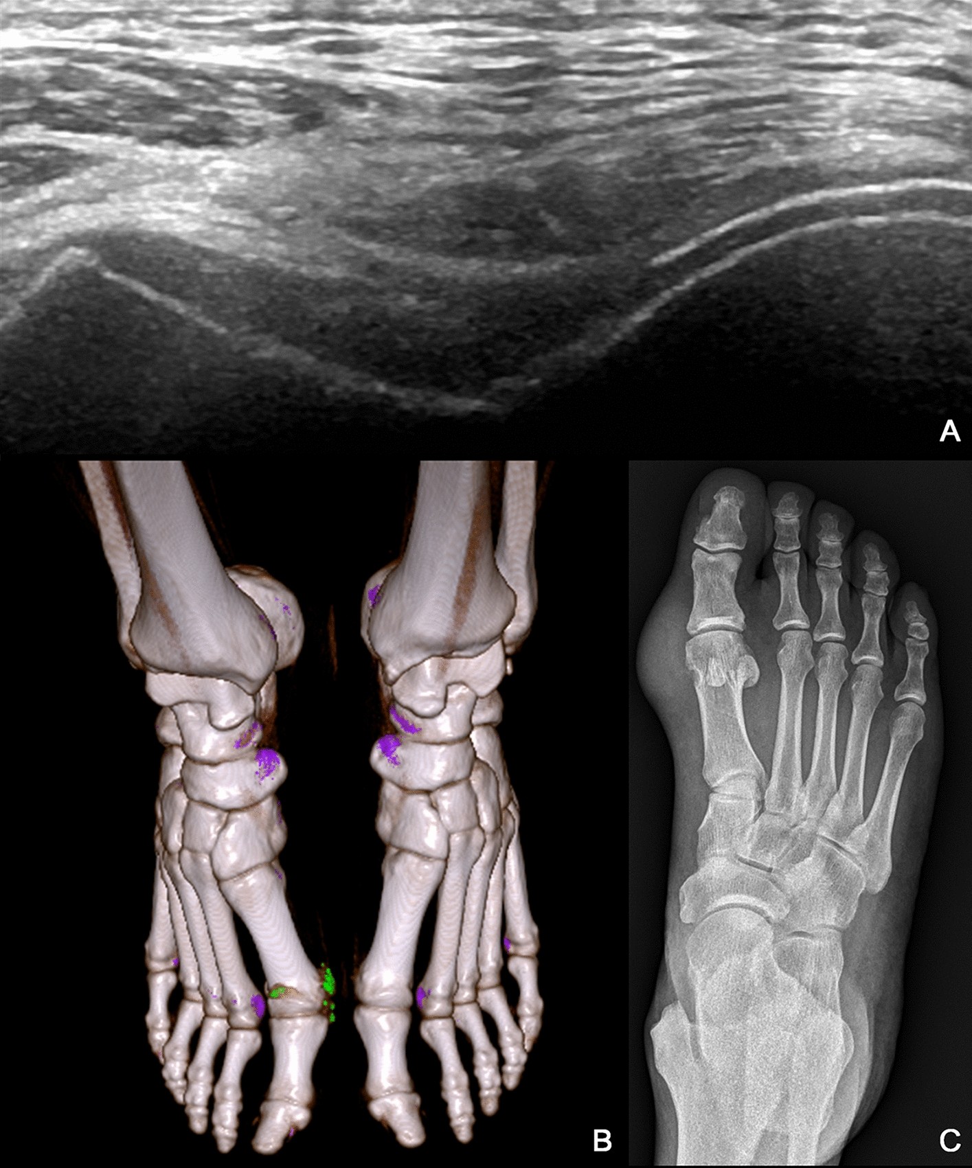
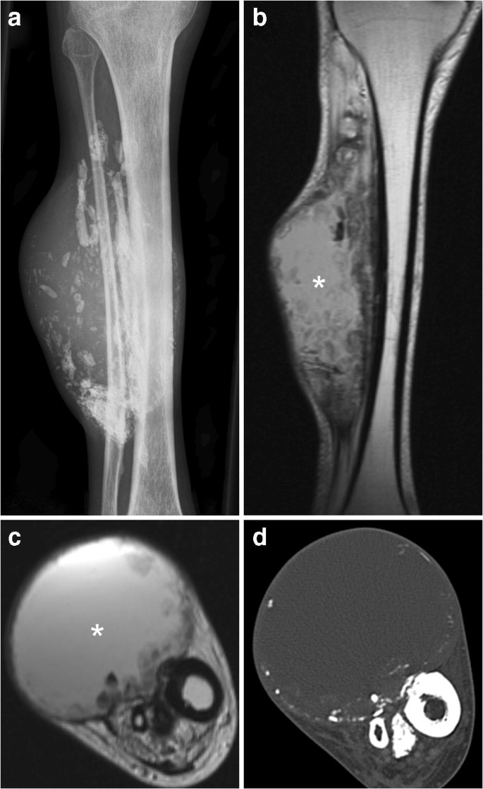
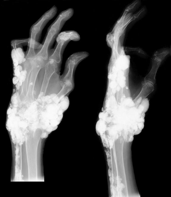
Abstract Musculoskeletal calcifications are frequent on radiographs and sometimes problematic. The goal of this article is to help radiologists to make the correct diagnosis when faced with an extraosseous musculoskeletal calcification. One should first differentiate a calcification from an ossification or a foreign body and then locate the calcification correctly. Each location has a specific short differential diagnosis, with minimal further investigation necessary. Intra-tendon calcifications are most frequently associated with hydroxyapatite deposition disease (HADD). In most cases, intra-articular calcifications are caused by calcium pyrophosphate dihydrate (CPPD) crystal deposition disease. Soft tissue calcification can be caused by secondary tumoural calcinosis from renal insufficiency, or collagen vascular diseases and by vascular calcifications, either arterial or venous (phlebolith). Teaching Points • Calcifications have to be differentiated form ossification and foreign body. • A musculoskeletal MRI study must always be correlated with a radiograph. • The clinical manifestations of calcifications may sometimes mimic septic arthritis or sarcoma. • HADD and CPPD crystal deposition have a distinct appearance on radiograph. • Calcinosis is more frequently caused by chronic renal failure and scleroderma.
Identification of tophi in ultrasound imaging based on transfer learning and clinical practice

Calcium Hydroxyapatite Deposition Disease - Sports Medicine Review
Calcified or ossified benign soft tissue lesions that may simulate malignancy
Soft Tissue Calcifications

Incidental findings in and around the prostate on prostate MRI: a pictorial review

A Compartmental Approach to the Radiographic Evaluation of Soft-Tissue Calcifications - ScienceDirect
Radiological identification and analysis of soft tissue musculoskeletal calcifications, Insights into Imaging

Dermatomyositis presenting with diffuse calcinosis
Magnetic resonance imaging of rheumatological diseases
Calcified or ossified benign soft tissue lesions that may simulate malignancy
Calcified or ossified benign soft tissue lesions that may simulate malignancy

A Compartmental Approach to the Radiographic Evaluation of Soft-Tissue Calcifications - ScienceDirect

Chapitre 17 – Tumeurs des tissus mous
de
por adulto (o preço varia de acordo com o tamanho do grupo)